In the ongoing battle against cancer, AI Medical service has been able to develop the world’s first gastric cancer differential AI that can help detect GI cancers at an early stage, potentially saving thousands of lives
Over the last 25-30 years, Japan has seen the rise of regional competitors who have replicated Japanese processes but doing so at a cheaper labor cost, pushing Japan out of mass markets. However, Japan is still a leader when it comes to the medical field. It has a 90% global market share in flexible endoscopes and a third of MRI systems around the world are made by Japanese firms. How have Japanese firms been able to maintain this lead in the medical field despite the stiff price competition?
Endoscopy equipment was originally developed in Japan, and even today there are more specialized doctors in this field than in peer countries. For example, the Japanese Gastroenterological Society is one of the world’s largest societies for endoscopic medicine and has more than 35,000 physician members. Japanese medical societies are involved in endoscopic medical activity worldwide. More than just manufacturing, medical advances require testing and developing devices. In Japan, there are opportunities to not only produce devices but also to test them in the field given Japanese expertise and the distribution of certain cancers present in Japan compared to other areas of the world. Furthermore, Japan has a national screening system for digestive tract cancers that utilizes endoscopic examination, so these exams are routinely performed. For example, in Saitama City, citizens who are over 40 years old can receive endoscopy examinations every year at the cost of only JPY 1,000 or USD 8*. Fittingly, more than 15 million endoscopic cases are performed every year in Japan.
*The amount varies depending on the municipality.
Japan's power lies not only in the number of endoscopic examinations performed but also in the quality of equipment used and the skill of those performing the exams. Besides machines or devices, human resources in the field of endoscopy are abundant in Japan. Furthermore, the Japanese endoscopic market remains a world leader since the development of endoscopic medicine 70 years ago. Besides the devices, I want to emphasize the respected ability of Japan's endoscopic medical associations and practitioners. Japanese doctors go overseas to teach about endoscopic examination to countries like Mexico, Brazil and Singapore. Endoscopists from Japan are very influential within the field. Endoscopic medical societies also support endoscopic manufacturers in improving product quality and making revisions to products. Japanese doctors help develop endoscopic techniques like microscopic endoscopy, colonoscopy, endoscopic mucosal resection, etc. To date, new endoscopic techniques have mostly arisen from Japan.
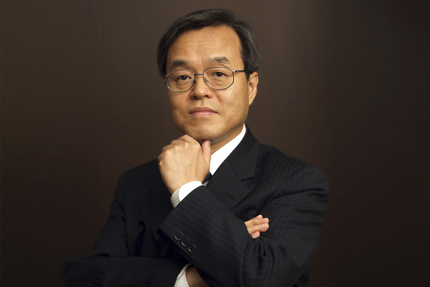
Can you give us an overview of your background, and how your expertise helps with the development of AI Medical's products?
I was a gastroenterological surgeon for five years at the beginning of my career, and I got my board certification from the Japanese Surgical Society. After that, I went to graduate school at the University of Tokyo where I studied for 4 years. I did my research on gene methylation as it relates to colorectal cancer. I obtained my MD and then my PhD. Around the time of my doctorate studies, a complete reading of the human genome was completed and genomics research was very popular. I researched some of the mechanisms by which cancer can arise based on genetic factors, particularly concerning colorectal cancer. Unfortunately, my investigations did not produce any results that could be used in the fight against colorectal cancer.
After graduating, I started my own clinic focusing on endoscopic examination for the gastrointestinal tract because the endoscopic exam is the only form of exam that can be used to detect early gastric cancer, esophageal cancer, and early colon cancer. No other medical examination can detect gastrointestinal cancers at an early stage as accurately. Of course, CT or MRI can be used to detect advanced cancers, but this is less clinically meaningful than early detection. Specifically, one of the issues with cancer, in general, is the degree to which the disease stage impacts survival and the quality of life. Early stages tend to have higher survival rates compared to later stages on the typical 5 or 10-year timescale. In particular, with some of the early cancers of the GI tract, there can be quite good 5-year survival rates and quality of life following resection. In the case of gastric cancer, 5-year survival remains 97% or 95% if the cancer is found at early stages, but just like any other cancer, things become much worse as the cancer progresses to stage three or four.
For stage zero or stage one stomach cancer, curative therapy is possible by removing a small amount of tissue. However, for gastric cancers of more advanced stages, two-thirds of or even the entire stomach has to be removed. Ignoring the impact of chemotherapy, radiation therapy or any other forms of treatment, removal of the stomach alone would ruin one’s relationship with food, sometimes causing severe weight loss and other issues. Doctors and patients would like to avoid that and try to keep cancer cases confined to the early stages, not advancing to later stages. It is vital for patients to do so. Japan’s endoscopic technology and techniques are extremely important to stay on that ‘early’ side of the early stage-late stage boundary.
15 years ago, an endoscopic examination was a difficult procedure for patients to undergo. It was not something they wanted to experience. Some of the techniques used at that time were focused on the diagnostic work itself and not so much on the patient’s experience. There was not much consideration of how willing or comfortable the patient would be with undergoing the procedure.
When I started my clinic 15 years ago, I wanted to concentrate more on sympathizing with the patients and understanding their perspective during the procedure, not just focusing on the diagnostics itself. I developed certain techniques that other facilities in Japan have begun to adopt. Patients do not feel as much resistance to endoscopic exams as before; they do not feel that it is as difficult physically, emotionally or spiritually as it used to be to undergo an endoscopic exam. Furthermore, there have been developments in endoscopic equipment. The diameter of the scopes becoming smaller, thinner, and finer contributes to the patient's comfort. We also have advances in anesthesia and other pre-exam preparation techniques.
My clinic gives more attention to the patient’s perspective in providing high-volume care. Overall, there are more than 8,000 endoscopic examinations done at my clinic each year. We have been able to perform those tests safely without adverse events while also providing good outcomes to the patients. We feel that worldwide adoption of Japan’s approach to gastrointestinal cancers will have positive effects. This approach includes a combination of advanced technology, therapeutic advances, and outreach for high-risk patients will enable other countries to discover most gastric cancers as is the case in Japan.
For example, in Japan, more than half of the procedures which are undertaken to remove gastrointestinal cancers are done in an endoscopic exam setting. We use tools for endoscopic exams rather than conducting major surgery, where patients will have to go through long hospitalizations and other burdens that we do not want the patients to face. One of the reasons why many Japanese physicians go abroad to spread Japanese medicine is that they developed the techniques to identify, remove, and treat cancers in this minimally invasive fashion; care providers in other nations are relatively unfamiliar with these techniques.
Gastric cancer approximately accounts for 30% of cancer deaths worldwide. It is estimated that up to 25.8% of early gastric cancers are overlooked because it is difficult to detect them early on. In 2018 you succeeded in building the world's first gastric cancer AI that can even detect early-stage cancers sometimes overlooked by endoscopic specialists. Why is it difficult to detect gastric cancers early on? What were some of the challenges that you had to overcome during this AI's development?
Let us assume that the mucosa of the stomach and the mucosa of the colon, different parts of the body, are both nice and flat. Before one has colorectal cancer, there is usually a kind of three-dimensional polyp that first forms like a mushroom in the large intestine (i.e., the colon). A polyp itself is not cancer, but polyps can undergo cancerous transformation. Most neoplastic lesions of the large intestine manifest as this type of polyp structure and are relatively easy to see during an endoscopic exam. In contrast to that, within the stomach the most common type of cancer is flat at the precancerous or early cancer stages; there is no three-dimensional polyp structure. Furthermore, the most common reason why cancer arises in the stomach is because of a long-standing inflammatory process. This inflammation is most likely due to H. pylori infection, which makes the mucosal tissue look red. Unfortunately, the cancer that arises in these high-risk areas is also rather red from inflammation. Hence, there is some similarity in terms of visual features between the cancer and the background.
As a result of this, unlike in the large intestine where we first have the polyp arising and then that easy-to-see structure becomes cancerous, with gastric cancer we have a flat lesion that blends in with the surrounding mucosa. In other words, it is more difficult to see early gastric cancer because it does not stand out, and it is not something that the human eye catches as easily. That is why up to 26% of early gastric cancers are overlooked during endoscopic examinations. With gastric cancer, advanced stage gastric cancer is easy for endoscopists to detect because there is a big, malformed serrated mass. However, detecting gastric cancer at its advanced stages (stage three or four) means more than half of the patients cannot be saved. Thus, we must detect gastric cancer at an early stage even if this is more difficult.
For example, out of 67 doctor participants, only one could correctly detect early gastric cancer among some of the images we presented to them that contained gastric cancer. In other words, more than 90% of doctors diagnose some cancer-containing images as normal.
This is the kind of reading test that we did to validate the product. We showed the AI images taken during endoscopic procedures and tested whether the AI would accurately locate the lesion or not. We showed that same image to doctors and asked them if they saw something strange or concerning in the image. The majority of doctors determined that there were no issues in some of the images we showed them, but there were lesions that should have been diagnosed.
In contrast, our AI system can detect almost all early gastric cancers. In this article, we demonstrated that our AI system overcomes Japanese-certified endoscopists based on sensitivity, or lesion detection rate. This was a statistically significant result. We are confident that our products can be introduced into clinical practice once doctors receive the proper training. We think that providing this to doctors on a day-to-day basis would lead to decreasing the rate at which gastric cancers are overlooked. AI has computer vision and has some differences with human vision, but at the end of the day, both still analyze the same imagery. In other words, things that are going to be difficult for AI are going to be difficult for humans. Things that are easy for AI to see are going to be easy for humans to see as well.
The colorectal polyp usually juts out and has a three-dimensionality to it, making it easy for the human eye to see. That is why AI for colorectal polyp detection can be developed using only 2,000 training images of polyps. For precancerous and cancerous lesions, it requires more than three times the amount of data to produce similar performance. That was a challenge we faced; the problem’s overall degree of difficulty is something that AI still has to overcome.
Our company could only overcome that challenge with lots of data. We utilized significant trial-and-error around the collection of the data. This includes assurances around data quality and the means by which data is collected, as well as how it is stored and analyzed. The entire data collection process involves making sure that the data is finely tuned to produce data that can create a high-performing AI. This was something that we had to figure out over years of trial and error. For example, when we receive data to develop AI for gastric cancer, we cannot just show that image to the AI. We have to label all data with certain features, and repeat this process for not just lesions; we also have to teach the AI what to ignore, including benign lesions like ulcers, inflammation or other issues.
We teach the AI what to pay attention to with certain types of labeling. This is manual labor that involves annotation by hand. It takes a lot of time to label each image by hand. We developed an in-house, proprietary annotation tool to assist in this task. Data collection and storage are also made more difficult by privacy concerns. Since privacy is a big issue in the medical field, anonymizing the data at the source at the earliest stage, and then using the proper storage and transmission methods after that were essential. The ideal form by which we get the data from our collaborative research partners is high-quality videos because these allow us to look at things frame by frame and find the best part to teach our AI. We consider what frames will help create the best AI. We have to watch the video and understand the meaningful parts and identify where the lesion is and determine the distance or type of lesions that we do not have a lot of in our teaching data. Adjusting and fine-tuning how you take still images out of the video to be shown to the AI is also a part of the data annotation process.
We advanced our collection methods based on the technology available in some cases and according to what we learned to be necessary to create good AI. We tried to refine those processes over time. For example, in 2017 when we just started our AI research, we only collected still images. However, after 2018, we started collecting high-definition data videos, and we gathered uncompressed videos from 2020 onward.
For something novel like our products, there is not a whole lot of precedent for what you are trying to achieve as well as the tools or means by which you are trying to achieve it. Due to not having things to reference or compare to, evaluating each step of the process is something that has to be done internally. The internal creation, validation and iterative process can be sometimes difficult, especially when you do not have outside resources or outside precedent examples to use.
As I mentioned, the H. pylori bacteria is the infection that leads to chronic inflammation, which then leads to the development of cancer. The first endoscopic AI that we put a lot of energy into was an AI that determines whether a patient had been infected with H. pylori or not. This was something that was not so difficult to develop and did not have as much clinical significance as an AI that can find cancer. Therefore, after achieving significant results with an AI for H. pylori, we decided to focus more on cancer products. Finding cancer can save lives more directly. We worked with our institutional partners in this way, to figure out what would be the most clinically significant and what type of products can provide value to patients and physicians.
What is this AI's success rate of detection when it comes to these gastric cancer lesions?
In most of the studies, it exceeds 90% sensitivity in detecting cancer lesions. This sensitivity metric seems to be the benchmark by which something is viewed as clinically meaningful. In the past, our AI demonstrated 94.1% sensitivity in our validation data (64 of 68 cases). Many of these cancers were tiny early-stage gastric cancers that even experienced endoscopists can miss. AI can assist in identifying suspicious cancer lesions more easily than before. I believe that with this AI, overlooked cancers will decrease significantly.
Partnerships have played a large role in accumulating data. You have done a good deal of joint research with academic institutions in Singapore. You also worked together with the University of Tokyo Hospital. Can you tell us more about the joint research that you have conducted with these institutions? Are you looking for similar opportunities in overseas markets?
My connection to the University of Tokyo goes back to when I graduated and took my PhD there. Afterwards, I was an endoscopist for more than 25 years. What differentiates our company from our competitors is that I, and many others, whom we have brought onto our team have a similar academic or clinical background. This experience puts us in a position to conduct good research. In addition, our academic and clinical experiences have allowed us to create a nice balance between business and research.
We are not focused on quickly turning our research into a product that we can sell, but we want our work and research to be turned into meaningful publications that will expand academic conversations about these topics. Many research partners are attracted to working with us because of our initial success and expertise. This is important overseas, where there is perhaps less awareness about gastric cancer due to having fewer cases or less experience and expertise.
In addition to working with the University of Tokyo, we have also worked with many domestic and overseas medical institutions including the National University Hospital of Singapore (NUH), the University of Hong Kong, Hospices Civils de Lyon, France as well as many other top-tier medical institutions in the United States. With some of these institutions, we are currently negotiating contracts to conduct collaborative research. With other institutions, we already have contracts and are just working out the details before we begin research activities. For others, collaborative research has been completed. For example, our study with NUH in Singapore has either been published or will be published soon. With this collaborative research, we can acquire foreign endoscopic imaging data and validate our AI system. For example, Japanese endoscopic tools are very advanced, and the top Japanese clinics have the newest equipment. This might not be the case even at top medical centers in the United States, because they do not have access to the newest equipment from manufacturers like Olympus and Fujifilm. We must respond to the needs of other markets to increase the diversity and representatives of our data set for our AI.
This technology is applied to the detection of gastric cancers. However, it is not only cancer that is difficult to detect in its early stages. There are other forms of cancer, such as pancreatic, kidney and liver cancers. Like gastric cancer, finding these early on can mean the difference between life and death. Are you looking to expand this technology to other forms of cancer?
Currently, we just focus on developing endoscopic AI. Other than gastric cancer, we are conducting research and development for esophageal cancer, which is also hard to detect in its early stage like gastric cancer. We are confident that our AI system can detect esophageal cancer in its early stages. Esophageal cancer can be divided into two main types histologically; squamous cell carcinoma and adenocarcinoma. In the United States and Europe, esophageal squamous cell carcinoma is not so common, but adenocarcinoma is. In Asian countries, the pattern is reversed, with more esophageal squamous cell carcinoma. This difference between the West and Asia is due to lifestyle and genetic factors. We are now developing an AI for esophageal squamous cell carcinoma and have also begun collecting data for esophageal adenocarcinoma.
According to the International Agency for Research on Cancer (IARC), before COVID-19, there were 17 million new cancer cases worldwide and 9.5 million deaths. They predicted that by 2040, there will be 27.5 million new cancer cases and 16.3 million deaths. Furthermore, we are seeing a dramatic increase in cancer cases for those 50 years of age and under. Between 2018 and 2021, cancer deaths increased by 4.7%. Why do you think we are seeing a global increase in cancer, and what do you think can be done to reduce this?
Multiple factors can cause cancer, and it is hard to isolate and say which one is the biggest contributing factor to increased cases. One reason may be the progress of diagnostic tools such as computed tomography (CT) and endoscopy. Endoscopic examinations are the only exams that can also detect the early stages of gastrointestinal cancers and aid in early treatment. The problem is that doctors overlook some cancers during endoscopic examination, and there are not enough qualified endoscopists in many countries. If gastrointestinal cancers are detected in their early stages, several million patients can be saved around the world.
Furthermore, as the world population grows yearly, more people are entering the age range during which their cancer risk is higher. In the developing world, people are now living beyond lung disease caused by pollutants or accidents that happen in the workplace; as populations are shifting from field or factory environments to office environments, we are seeing overall improved health outcomes outside of cancer. Unfortunately, older populations tend to have more cancer. If people survive into their 70s, they are in the age range that is predisposed to cancer. Having a high number of cancers is never desirable, but it also speaks to the accomplishments that have been achieved in public health in different nations.
Japan has the oldest society in the world and we know that roughly 50% of cancer occurs in people who are over the age of 65. Older age can mean different biological differences in tumor or microenvironment behaviors. Along with Japan's aging population, there is also a labor crisis which makes it a challenge to pass on the knowledge you have accumulated over many years. What are some of the challenges and opportunities this demographic shift has presented for AI medical Service?
Older people are going to have more diseases and will be more at risk for cancer. Being older, their bodies might not be able to withstand major surgeries to remove tumors or organs or chemotherapy. Providing solutions that can address their needs and diagnosing their cancer at early stages can help avoid the need for late-stage therapies that they might not be able to withstand due to their advanced age. With gastric cancer in Japan, more than half of them are now removed through endoscopic procedures (not major surgeries).
The Japanese medical field is constantly investigating fundamental workflows about approaching cancer in terms of how it is diagnosed and treated, and we hope this figure can improve up to 70-90%. We think that this is possible to achieve even with the aging society.
Furthermore, Japan has more endoscopists per capita than any other nation, but many of these experienced doctors are aging as well. Over the age of 50, there are physical factors due to aging that can affect doctors' abilities to carry on their work, such as deteriorating vision. Using AI and other tools renowned for vision or ability to see certain features and combining that with their expertise and ability to manage their patients is a good way of extending doctors' careers. We hope that this can stave off the problem of physician understaffing and overworking in Japanese medicine. This also applies overseas, where there are fewer doctors per capita.
Of course, the purpose of AI is not to replace endoscopists. We just want to make their work easier and with many aging doctors, AI can help them have a longer career in medicine. The average age in this field in Japan is over 60. We are thinking of introducing our products into the market over the next 5-10 years, potentially contributing to endoscopists working 5-10 years more should they want to do so. This was the opportunity presented to our company by Japan's aging society.
In 2022, you opened operations in the United States and Singapore. Last month, you opened a new office in New York as well. Can you tell us what your expectations are for these regions moving forward, and have you identified any other countries that you would like to expand into?
Most gastric cancers in the United States are in the advanced stages. Partly because of the low incidence of gastric cancer, doctors in the US do not have enough opportunities to gain knowledge of and experience with cancer at an early stage. Furthermore, unlike in Japan, no screening program for gastric cancer is established in the US. Japan has a relatively high incidence of gastric cancer compared to other nations, but we have a low mortality rate because of having established screening programs and more endoscopists per capita. These endoscopists also undergo advanced training and use cutting-edge technologies. We hope to spread awareness and share some of our methods to detect gastric cancer at an earlier stage with the United States.
Gastric cancer has a very low incidence in the US, but there are still more than 25,000 cases. The cases are clustered especially on the east and west coasts with a large immigrant population, particularly from Northeast Asia (Japan and Korea). Gastric cancer is also seen among populations with lower socioeconomic status. That is because the likelihood of Helicobacter pylori infection, which leads to gastric cancer, is related to sanitation and socioeconomic status.
In the United States, it might not be worth screening the entire population, but some subpopulations warrant outreach. We think that we can work with these communities to help isolate and reach high-risk patients, while also providing endoscopists with the tools that they can use to help these patients. Early diagnosis requires a whole package, so we cannot do it alone. There needs to be a change in the process and awareness overseas. We hope to help drive that change.
For example, Stanford Medicine has a program called the Center for Asian Health Research and Education (CARE). It is about reaching out to Asian communities and populations and providing solutions that are specific to their needs, particularly people from Northeast Asia or Asia in general. When they move to the United States, they retain their gastric cancer risk for generations. It is not something that drops off immediately. However, due to the local health system being geared more toward the needs of Caucasian patients, there could be a gap in meeting Asian patients' needs. We thought it would be great to collaborate with CARES. New York is another great hub for innovation and other issues. Collaboration is important for our outreach efforts because of their patient diversity and expertise. They also have a Japanese doctor on their staff and Japanese researchers whom we can work with to ensure that things would move smoothly.
You founded the company in 2017 and have already achieved so much, including the development of endoscopic AI. If we come back in five years to interview you again, what would you like to have achieved by then?
Our AI system is on the premises and it involves a physical unit. We have one unit per endoscopy room and there are physical cable connections to the device that has to be set up. There is delivery, setup and maintenance involved. In the future, I am most excited about being able to share our AI easily through the cloud and turning the software into an online platform. In other words, we are thinking about removing the hardware component and shifting to something completely software and digital to expand overseas.
We have to overcome many things in terms of technology, communications and IT environment for the hospitals. Furthermore, we also must deal with local regulations not only in terms of government but also hospitals regarding data sharing and privacy. Data security can be another issue. If we find solutions for these challenges, we can create a platform where all doctors can be connected through an AI platform. The goal is not just to consolidate AI or use AI services, but also for endoscopists to support each other in their diagnosis and treatment. We want to create a community for endoscopists no matter where they are, what tools they are using and whatever working environment they might have.
Are you planning on doing this In-house, or are you planning to collaborate with other companies?
We are flexible and feel that we can succeed with either approach based on our collective skills and experience. I worked for the endoscopic medical society for more than 25 years, and I have always contributed to the advancement of endoscopic medicine. I developed and systematized a painless endoscopic insertion technique that prevents bowel perforation. Likewise, I also developed and introduced a ‘cold polypectomy’ technique that reduces the risk of complications following colon polypectomy to almost zero.
I am also the owner and operator of my own clinic which is now considered one of the top clinics specializing in gastrointestinal endoscopy in Japan. Lastly, I started the company five years ago focusing on research and development of endoscopic AI because our mission is to eliminate gastroenterological cancers and save many patients around the world. In the future, I would like to create a connected platform for doctors worldwide to collaborate in real-time. I want to contribute to the evolution of endoscopic care worldwide and contribute to the future of endoscopic medicine.
Interview conducted by Karune Walker & Ana Ruiz


0 COMMENTS